CategoriesRemedial Massage/Myotherapy

Using Myotherapy to Exercise More Efficiently

Using Myotherapy to Exercise More Efficiently

What Is Myotherapy?

Myotherapy is a great way to reduce muscle tension, increase range of motion and assist with pain reduction. It is important to know why we are in pain, what caused it and how to prevent the same pain/injury from occurring again. Research has shown that adopting a multimodality method to assist with complex injuries consistently provided significant improvements in pain and function. These modalities may include Doctors, Specialists, Physiotherapists, Myotherapist, and Remedial Massage therapists all working together to manage you as a client. Further benefits of working in a multi-disciplinary approach include improved communications between the healthcare professionals and guarantees that we are working holistically (i.e. taking a whole-body approach to healthcare), which research indicates achieves the best outcomes for the patient. 

Exercise is vital for rehabilitating the body and preventing injuries however, it can be difficult for our body to work efficiently if we are in pain. Our brain is very smart in trying to reduce painful movements when injured so it will do anything it can by using compensatory patterns to still achieve movement in pain. Therefore, you might even feel tight and sore on the other side of the injury due to the body compensating to reduce the stress on the injured site.  

Myotherapy and massage can be an excellent treatment to address the changes in the body as a result of injuries and manage the tissues around the site of the injury to ensure that the body is working efficiently which will increase the effectiveness of rehabilitation exercises.     

Written by Mo Bhatnagar (Clinical Myotherapist) 

References:  
    1. Peterson, K., Anderson, J., Bourne, D., Mackey, K., & Helfand, M. (2018). Effectiveness of Models Used to Deliver Multimodal Care for Chronic Musculoskeletal Pain: a Rapid Evidence Review. Journal Of General Internal Medicine, 33(S1), 71-81. https://www.ncbi.nlm.nih.gov/pmc/articles/PMC5902347/ 
    2. Jonas Gopez, M. (2021). Exercise and Back Pain. Retrieved 4 May 2021, from https://www.spine-health.com/wellness/exercise/exercise-and-back-pain  
CategoriesMedical Condition

Tennis Elbow

Tennis Elbow

What is tennis elbow? 

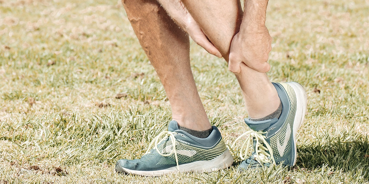
Tennis elbow, also known as lateral epicondylitis, is a common painful condition of the elbow that affects between one to three people in every 100. The condition is a type of tendinopathy and typically presents itself as inflammation and pain occurring on and around the lateral epicondyle, a bony bump on the outside of the elbow, where the tendons attach to the bone. 


This condition most commonly affects individuals between the ages of 30 and 50 years old, but it can occur in all ages, in both men and women, and in both arms (however the dominant arm is usually more prevalent). 

The condition usually develops gradually and, depending on how severe it is, can last between six months and two years. 

What are the symptoms? 

    • Pain and tenderness on the outer side of the elbow and at times, the pain may even travel down the forearm.  
    • Pain and/or weakness with gripping, writing, and twisting movements of the forearm, as well as lifting and carrying items. 
    • Pain when the forearm muscles are stretched and tender spots in the forearm muscles.  
    • In some cases, neck stiffness and tenderness, and signs of nerve irritation like numbness and pins and needles. 

What causes it? 

Despite its name, most people don’t get it from playing tennis. Sports such as tennis are commonly associated with this condition, but the problem can occur with many different activities. Tennis elbow is thought to occur due to mal-adaptations to the tendon. This is often caused by overloading the tendon through repetitive gripping and grasping activities such as hammering, painting, and typing. Other contributing factors may include unaccustomed hand use, weak forearm muscle strength or tight muscles, and/or poor technique. 

Although less common, a direct blow to the elbow may result in swelling of the tendon that can lead to degeneration. This can make the elbow more susceptible to an overuse injury. 

How is tennis elbow diagnosed? 

Your physiotherapist or doctor can clinically diagnose your tennis elbow. After obtaining a detailed history and performing some special tests, they may determine a provisional diagnosis of tennis elbow. It is important that your physiotherapist also assess your neck and upper limb neurodynamic as referred pain from the neck and reduced nerve mobility can mimic tennis elbow. Failure to do so may result in a lack of symptom improvement. 

An ultrasound scan or MRI are the best tests to identify tendon changes and adaptations, but are often not necessary. X-rays are of little diagnostic benefit. 

Managing your symptoms 

Most people who have tennis elbow find that their symptoms get better when they rest their arm and take-over-the counter pain medications. If you do this and still have symptoms after six weeks or so, visit your GP or physiotherapist. 

Generally, the most helpful approach is to implement strategies that reduces pain. This may include changing the way you are doing things.  

Ways to try to reduce the pain 

    • Rest your elbow and arm as much as you can. Avoid or modify activities and movements that make your pain worse. If you need to lift something heavy, bend your elbows and make sure the palms of your hands are facing upwards. 
    • Use anti-inflammatory, non-steroidal or ‘cold’ gels or creams, which can be rubbed into the painful area. 
    • Apply an icepack or a bag of frozen peas wrapped in a tea towel on your elbow may temporarily relieve the pain. Avoid putting the ice pack directly onto your skin to avoid causing damage to your skin. Only use it for about 20 minutes every three to four hours. 
    • Stretches can also give relief. 
    • Use a tennis elbow brace. You may find this is helpful to “offload” the area and allow you to continue with normal activities. 

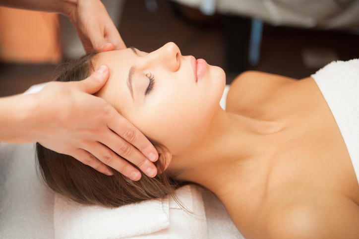
The role of physiotherapy 

Physiotherapy has been shown to be effective and helpful in the short and long-term management of tennis elbow. 

Physiotherapy aims to:

    • Reduce elbow pain. 
    • Facilitate tissue repair. 
    • Restore normal joint range of motion and function. 
    • Restore and improve muscle length, strength and movement patterns. 
    • Normalise neck function and upper limb neurodynamic. 

These goals can be achieved in various ways and, following a detailed assessment of your elbow, arm and neck, your physiotherapist can discuss the best strategy for you to implement based on your symptoms and your lifestyle. 

Physiotherapy treatment may include gentle mobilisation of your neck and elbow joints. Other treatment options available include elbow taping, muscle stretches, neural mobilisations, massage and strengthening. 

If you think you have tennis elbow come visit us at Elevate Physio and Pilates in Balwyn for an initial assessment, where we provide a comprehensive evaluation, explain the diagnosis, and highlight the different management options! 

Written by Mo Bhatnagar (Clinical Myotherapist) 

CategoriesMedical Condition

Understanding the Shoulder Joint

Understanding the Shoulder Joint

The Shoulder is a complex collection of 4 joint in the body Based on the amount of movements required, the shoulder is naturally an unstable joint hence why injuries around the shoulders are very common.

Shoulder Anatomy and relation to shoulder injuries

The Shoulder is a complex collection of 4 joint in the body, these include: 

  • Acromioclavicular joint (AC Joint)
  • Sternoclavicular Joint (SC joint) 
  • Glenohumeral Joint (GH Joint) 
  • Scapulothoracic Articulation

Based on the amount of movements required, the shoulder is naturally an unstable joint hence why injuries around the shoulders are very common. Common injuries we typically see include rotator cuff injuries, shoulder impingement, sub-acromial bursitis, frozen shoulder (Adhesive capsulitis), and dislocations from contact sports and falls. The shoulder is heavily reliant on the muscles to provide stability in the motions that the body requires. Therefore, the shoulder needs to have adequate strength through the muscles to reduce muscular bias which can pull the shoulders away from the appropriate position and place unnecessary stress on other structures which can cause and exacerbate injuries and pain. 

Shoulder pain and the role of physiotherapy/myotherapy

As the shoulder is comprised of many joints and muscles to provide movement. Pain and dysfunction of the shoulder is usually multifactorial meaning there may be a collection of the joints and muscles which are causing the problem. Your Physiotherapist/Myotherapist may need to assess multiple areas of the shoulder, in different positions and ranges to identify the most likely causations and risk factors for the shoulder pain based on your sport, work, and daily activities. We then use the information to create a plan on what muscles may need releasing through manual therapy and stretching, and which muscles need strengthening to reposition the shoulder and improve the function which may be causing pain. 

Treatment for shoulder conditions can vary based on age, previous history, and daily activities. A combination of exercise intervention and manual therapy such as soft tissue massage, dry needling, and stretching techniques have shown to be very effective in increasing strength, decreasing pain, as well as improving the range and function of the shoulder. 

Written by Mo Bhatnagar BHSc. (Clinical Myotherapy) 

References  
    1. Go, S., & Lee, B. (2016). Effects of manual therapy on shoulder pain in office workers. Journal of Physical Therapy Science, 28(9), 2422-2425. doi: 10.1589/jpts.28.2422
    2. Pieters, L., Lewis, J., Kuppens, K., Jochems, J., Bruijstens, T., Joossens, L., & Struyf, F. (2020). An Update of Systematic Reviews Examining the Effectiveness of Conservative Physical Therapy Interventions for Subacromial Shoulder Pain. Journal Of Orthopaedic & Sports Physical Therapy, 50(3), 131-141. doi: 10.2519/jospt.2020.8498  
CategoriesMedical Condition

Peripheral Nerve Entrapment

Peripheral Nerve Entrapment  

What is Peripheral Nerve Entrapment? 

Peripheral Nerve entrapment (PNE) is a common problem in the adult population. These can affect both the upper body in the arms and fingers and the lower body into the legs and feet. For the upper body, PNE can usually cause common conditions such as carpel tunnel syndrome, cervical disc bulge, and thoracic outlet syndrome whilst lower body entrapment may include lumbar disc bulge, piriformis syndrome, and peroneal nerve entrapment.  

What are the symptoms? 

The symptoms of PNE are usually similar between the upper and lower extremities with tingling, numbness, and sometimes loss of function which can range from mild to severe weakness. Neural pain can usually be described as burning, sharp, and shooting pain down the arms or legs. 

How can peripheral nerves cause pain and dysfunction? 

Due to the intricate pathways some nerves take, they can be susceptible to irritation or damage due to compression or friction caused by external sources such as prolonged sitting or direct impact such as hitting the ‘funny bone’ which isn’t an actual bone, but rather an exposed nerve that reacts with impact. Internally these nerves can be affected by compression and friction from various structures that surround the nerve such as bones and muscles. Long-lasting compression or friction can result in compromised blood flow to the nerve as well as inflammation around the nerve. 

Difference between muscle referral and Nerve Pain? 

Muscle referral or trigger point pain and nerve pain can feel similar with pain usually traveling down to an extremity, except nerve pain will usually accompany an altered sensory (tingling, numbness) or motor (weakness) symptom which is different to a muscle that may feel like more of a dull ache, soreness and may increase symptoms with pressing on the affected area. Neural pain is usually only worsened by putting the nerve on stretch or by sitting or lying in a certain position that may increase the numbness and tingling sensations. Health professionals such as Myotherapist and Physiotherapists use palpation, movements, and orthopaedic tests that help in determining the cause and area of where the nerve may be causing entrapment issues. 

How is neural pain treated?  

As Myotherapists we firstly use our extensive knowledge of anatomy and structures to identify what nerve is being entrapped and follow that to what spinal level that nerve will originate from. From there we can perform special tests to identify whether the problem is within the spine or elsewhere. These tests can include strength tests, stretch tests, and muscle palpations to identify the type of entrapment and where it may originate from. Once the nerve is identified we can use techniques such as soft tissue muscular massage, dry needling, and Transcutaneous Electrical Neural Stimulation (TENS) work to relieve and free up the nerve from being entrapped. We will then accompany the treatment with some home exercises to assist the entrapped nerve, these may include muscle release work by a spikey ball or foam roller, neural floss techniques, and adjusting positions in sitting and sleeping to ensure symptom management. 

Nerve entrapment can be debilitating and cause major disruptions to a person’s day to day life therefore, it is important to try and get the symptoms under control as soon as possible to prevent the symptoms from becoming worse as well as future complications. 

Written By Mo Bhatnagar (BHSc. Clinical Myotherapy) 

References  
    1. Ulnar Nerve Entrapment at the Elbow (Cubital Tunnel Syndrome) – OrthoInfo – AAOS. (2021). Retrieved 11 May 2021, from https://orthoinfo.aaos.org/en/diseases–conditions/ulnar-nerve-entrapment-at-the-elbow-cubital-tunnel-syndrome  
    2. Nerve entrapment. (2021). Retrieved 11 May 2021, from https://www.physio-pedia.com/Nerve_entrapment
    3. Nerve Entrapment – Neurosurgical Associates of Central Jersey. (2021). Retrieved 11 May 2021, from https://neurosurgerycnj.com/peripheral-nerve/nerve-entrapment/  
CategoriesHow To

What’s Really the Correct Way to Lift Something?

Correct way to Lift

What Is The Correct Way to Lift Something?

For as long as we can remember, we have always been told that flexing your lumbar spine during lifting is dangerous and that it can lead to discs “slipping” or “blowing out”  

For as long as we can remember, we have always been told that flexing your lumbar spine during lifting is dangerous and that it can lead to discs “slipping” or “blowing out”. The common advice is to maintain a “neutral” spine, which is a specific position of the spine that is regarded as safe from injury, as seen in Figure 1. Whether this belief is true is still heavily debated, but the important thing to remember is that CONTEXT IS EVERYTHING.  

Image result for neutral spine

There is some argument that lumbar flexion, specifically repetitive flexion 1 and long duration stretch under load 2 may pose some risk for injury. A study has found that lifting heavy objects with lumbar flexion led to higher rates of compression on the lumbar spine compared to maintaining a neutral spine.3 However, this train of thought should be questioned, as a study comparing a neutral and flexed lumbar spine found that disc herniation was more prevalent in a neutral spine compared to a flexed one.4 Important to note is that these studies were completed in non-living people, meaning its results may not be applicable to you. This is an important consideration, as discs and any other living tissue of the body can adaptively heal and strengthen over time as the human body is extremely adaptable when exposed to the right stimulus. 

As aforementioned, context is everything when deciding whether lumbar flexion is dangerous when lifting. If you are lifting something that is low load, such as picking clothes off the floor, then it most likely does not matter whether the lumbar spine is flexed or neutral. This is because our spines love movement in any direction and was designed to be flexed. This is demonstrated by a recent systematic review which found that greater lumbar flexion during lifting was in fact NOT a risk factor for developing LBP, nor was it a differentiator of people with and without LBP.5 However, if lumbar flexion is irritating for you modification of how you lift may be warranted initially until your back is desensitised to the movement!  

If you are lifting heavy objects, such as a 20kg bag of cement or even a barbell, the general recommendation would be to lift with more of a neutral spine as there is a slightly higher predisposition of risk of injury with loaded lumbar flexion. However, even if you were to lift with a flexed lumbar spine it does not mean your disc is going to sustain an injury. Regardless of whether you lift with a neutral or flexed lumbar spine, if you progressively load the body and increase its tolerance to load then your risk of injury reduces as the body is extremely adaptable. Anecdotally, there are two types of clients we see who injure their backs from heavy lifting. The first are those who suddenly start lifting heavy objects without prior training/stimulus and have not progressively loaded their back. This may be individuals who have just started a new manual job and have had a period of relative rest or those new in the gym who have started lifting something too heavy too quickly. The second type of clients we see are those that injure their backs from “awkward” lifting techniques due to time constraints and work demands. This awkward technique has little to do with the fact of whether they had a neutral or flexed lumbar spine, but rather they overreached when lifting something heavy or did not have a stable base of support to initiate the lift. To ensure that your back is protected, the team at Elevate Physiotherapy & Pilates are perfectly placed to comprehensively assess risk factors for developing low back pain (LBP) and implement an exercise program to mitigate future episodes of LBP.   

In conclusion, with everyday tasks that involve low load lifting, it does not matter whether a neutral or flexed lumbar spine is maintained, but when lifting something heavy it is recommended to maintain a neutral spine as there is a slightly greater predisposition for injury. Whilst the jury is still not out in terms of whether lifting with a neutral spine is better than a flexed one, the important takeaway from this blog is that progressive loading of the back is the best method to reduce the likelihood of injury with heavy lifting.  

Want a lifting/ergonomic assessment? Call us 9836 1126.   

Written by Mo Bhatnagar (Clinical Myotherapist) 

References
    1. Callaghan, J. P., & McGill, S. M. (2001). Intervertebral disc herniation: studies on a porcine model exposed to highly repetitive flexion/extension motion with compressive force. Clinical Biomechanics, 16(1), 28-37. 
    2. Solomonow, M. (2012). Neuromuscular manifestations of viscoelastic tissue degradation following high and low risk repetitive lumbar flexion. Journal of Electromyography and Kinesiology, 22(2), 155-175. 
    3. Wade, K. R., Robertson, P. A., Thambyah, A., & Broom, N. D. (2014). How healthy discs herniate: a biomechanical and microstructural study investigating the combined effects of compression rate and flexion. Spine, 39(13), 1018-1028. 
    4. Veres, S. P., Robertson, P. A., & Broom, N. D. (2010). ISSLS prize winner: how loading rate influences disc failure mechanics: a microstructural assessment of internal disruption. Spine, 35(21), 1897-1908. 
    5. Saraceni, N., Kent, P., Ng, L., Campbell, A., Straker, L., & O’Sullivan, P. (2020). To flex or not to flex? is there a relationship between lumbar spine flexion during lifting and low back pain? A systematic review with meta-analysis. journal of orthopaedic & sports physical therapy, 50(3), 121-130. 
CategoriesMedical Condition

Imaging for Low Back Pain: More Harm Than Good?

Imaging for Low Back Pain: More Harm Than Good?

Getting imaging for low back pain (LBP) is very common practice, with 42% of clients with LBP receiving an X-ray, CT, or MRI within 1 year of diagnosis. While it may be intuitive to get imaging for an acute episode of LBP, there are several reasons why it may in fact cause more harm than good 

Firstly, what the research has found is that the “findings” on imaging are often not correlated with the symptoms of your LBP, meaning the “findings” are not likely causing your pain.2 Studies have found that if you scan healthy people with no LBP, there is a very high prevalence of abnormal image findings such as “spondylosis” and “degeneration”.2 This prevalence of abnormal image findings in a healthy population increases with age, 2 as seen in the Table 1. Consequently, imaging is not indicated for LBP in the absence of red flags, on the basis that the rate of abnormal imaging findings is high in healthy people and do not correlate well to your symptoms. 

Table 1. Prevalence of abnormal image findings in people with no low back pain (LBP).2 

Age of people with no LBP: 

Percentage with abnormal image findings: 

20 

37% 

30 

52% 

40 

68% 

50 

80% 

60 

88% 

70 

93% 

Secondly, the imaging results may be misinterpreted by clinicians and can result in unhelpful advice, needless subsequent investigation, and potentially even invasive interventions, such as surgery.1 whilst invasive interventions may be warranted in some cases with identified red flags, the vast majority of people with LBP can be managed non-surgically and can have equally as good outcomes.  

Thirdly and most importantly, misinterpretation of image findings, such as “degenerative disc disease”, “disc protrusion” and “foraminal narrowing”, can result in catastrophisation of the condition, which is defined as a person viewing their situation as considerably worse than it actually is. Catastrophising promotes an extremely unhelpful belief in people that their spines are structurally damaged and vulnerable.1 consequently, clients often become more fearful of movement and activity, and usually opt for bed rest. This contrasts with best practice for acute LBP, as bed rest and immobility are not recommended compared to staying active within pain limits, and that advice to stay active had greater benefits in pain relief and functional improvements.3 Clients misinterpreting their image results also lowers their expectations for recovery as a study found that the early use of MRI for LBP resulted in increased disability, poorer perceived prognosis and a greater chance of undergoing back surgery.1  

In conclusion, in the absence of red flags, imaging for low back pain is contraindicated as image findings do not correlate with symptoms and may promote detrimental beliefs about LBP that can worsen outcomes. The red flags which would warrant imaging for LBP are severe and/or progressive neurologic deficits, LBP that is constant and does not change with movement, and incident of trauma. Engaging with a health professional, such as a physiotherapist, will assist in ascertaining the presence of red flags and suitability for imaging. 

References
    1. Darlow, B., Forster, B. B., O’Sullivan, K., & O’Sullivan, P. (2017). It is time to stop causing harm with inappropriate imaging for low back pain. Brinjikji, W., Luetmer, P. H., Comstock, B., Bresnahan, B. W., Chen, L. E., Deyo, R. A., … & Jarvik, J. G. (2015). Systematic literature review of imaging features of spinal degeneration in asymptomatic populations. American Journal of Neuroradiology, 36(4), 811-816. 
    2. Dahm, K. T., Brurberg, K. G., Jamtvedt, G., & Hagen, K. B. (2010). Advice to rest in bed versus advice to stay active for acute low‐back pain and sciatica. Cochrane database of systematic reviews, (6).
CategoriesMedical Condition

Cervicogenic Headaches What Are They and How Can They Be Treated?

Cervicogenic Headaches What Are They and How Can They Be Treated?

A Cervicogenic headache is a “headache caused by a disorder of the cervical spine and its component bony, disc, and/or soft tissue elements, usually but not invariably accompanied by neck pain” 

What is a Cervicogenic headache?

According to the International Headache Society, a Cervicogenic headache is a “headache caused by a disorder of the cervical spine and its component bony, disc, and/or soft tissue elements, usually but not invariably accompanied by neck pain”.

What are the symptoms? 

Cervicogenic headaches typically start at the neck and base of the head and works its way up to the eye like a question marks, as seen in Figure 1 


Figure 1. Cervicogenic Headache Pain Pattern (Source: https://www.ccsbismarck.com/cervicogenicheadaches/) 

What causes it? 

Around the neck level, there are several nerves which innervates the structures on the upper neck that joins with a nerve that supplies structures at the front of the face, thus when the upper neck nerve is irritated it can cause the pain to travel to the front of the face.  

How is Cervicogenic headaches diagnosed? 

The major criteria which may indicate that it is indeed Cervicogenic headache includes: 

  • Restriction of neck range of motion. 2 
  • Pain or tenderness upon touch of the neck. 2 
  • Typically one-sided neck, shoulder or arm pain that does shift sides. In rare cases it may occur on both sides. 2 
  • Reproduction of symptoms by neck movement and sustained awkward head positioning.2 
  • Moderate to severe non-throbbing pain. 2 

Whilst these are some of the criteria to rule in a Cervicogenic headache, there is overlap between other types of headaches such as tension or migraines. Therefore, it is essential to get a health professional, such as a physiotherapist, to assist in differentiating the different types of headaches as the management strategies for each type is different.  Our physiotherapists at Elevate Physio and Pilates are exceptional at differentiating the different types of headaches through a thorough verbal and physical assessment.  

Managing your symptoms 

While it may be worrying to have the pain move towards to the front of the face, Cervicogenic headaches are not serious and can be exceptionally managed by physiotherapists. 

The Role of Physiotherapy 

Physiotherapy is considered the first-line treatment of Cervicogenic headaches. Physiotherapy treatment includes manual therapy around the neck and shoulders in conjunction with an exercise program. Research has found that a 6-week program of manipulative therapy and supervised exercise reduced the symptoms and frequency of Cervicogenic headaches, with the effects being maintained in the long-term.3 

If you think you have a Cervicogenic headache come and see us at Elevate Physio and Pilates in Balwyn for an initial assessment, where we provide a comprehensive evaluation, explain the diagnosis, and highlight the different management options! 

References 
  1. https://ichd-3.org/11-headache-or-facial-pain-attributed-to-disorder-of-the-cranium-neck-eyes-ears-nose-sinuses-teeth-mouth-or-other-facial-or-cervical-structure/11-2-headache-attributed-to-disorder-of-the-neck/11-2-1-cervicogenic-headache/#:~:text=Description%3A,invariably%20accompanied%20by%20neck%20pain. 
  2. Hall, T., Briffa, K., & Hopper, D. (2008). Clinical evaluation of cervicogenic headache: a clinical perspective. Journal of Manual & Manipulative Therapy, 16(2), 73-80. 
  3. Jull, G., Trott, P., Potter, H., Zito, G., Niere, K., Shirley, D., … & Richardson, C. (2002). A randomized controlled trial of exercise and manipulative therapy for cervicogenic headache. Spine, 27(17), 1835-1843. 
CategoriesRemedial Massage/Myotherapy

Can Massage Help With Relieving Stress?

Can Massage Help With Relieving Stress?

Today, most of our population lead a very busy lifestyle, which can lead to stressful times. In an ideal world, no one wants to be stressed, but sometimes events eventually catch up with us and it seems everything needs to be done at once which creates stress. Stress can be classified as physical, emotional, and mental. Each type of stress can have various effects on our bodies and wellbeing, so it is important to keep in check with mindful techniques such as meditation, yoga and even walking to assist during stressful periods. Some occupations are coined for their high-stress nature due to completing multiple tasks at a time for an extended period these include teachers, nurses, emergency services, office workers, and even students.

Is massage therapy effective?

Multiple studies have been conducted on the effects of massage therapy on students, patients under cardiac care, and high-stress occupations. All of these studies have all shown significant improvements in patient wellbeing from self-appointed stress and anxiety questionnaires, as well as a decrease in levels of cortisol (the stress hormone) and reduced heart rate/blood pressure with massage therapy.  

Many of these studies have shown the differences between massage therapy as compared to self-guided relaxation such as meditation tapes and standard care. With each study, massage therapy was concluded to have more significant results for stress and anxiety levels as compared to other forms of practice. The increase in effectiveness of massage therapy could be due to the effect on the neurological system that assists in relaxing the sensory receptors on the skin surface that connect to other areas of the body via neurons such as the muscles and in some cases organs. This helps to settle our ‘Flight or Flight’ response which increases our levels of cortisol and helps introduce our ‘rest and digest’ nervous system response as well as release endorphins. Other effects of massage include movement of blood flow and lymphatic fluid as well as providing heat and healing to the body which relaxes muscles tension and aids in tissue healing. 

If you are feeling constantly stressed, the effects of massage therapy in conjunction with a healthy lifestyle and adequate amount of exercises are the building blocks of leading a happier and overall healthier life. Call us today to book in 9836 1126.  

References
    • Zeitlin, D., Keller, S., Shiflett, S., Schleifer, S., & Bartlett, J. (2000). Immunological Effects of Massage Therapy during Academic Stress. Psychosomatic Medicine, 62(1), 83-84. 
    • Yousefi, H., Mirzamohamadi, M., & Nazari, F. (2015). The effect of massage therapy on occupational stress of Intensive Care Unit nurses. Iranian Journal of Nursing and Midwifery Research, 20(4), 508.  
    • Sharpe, P., Williams, H., Granner, M., & Hussey, J. (2007). A randomised study of the effects of massage therapy compared to guided relaxation on well-being and stress perception among older adults. Complementary Therapies in Medicine, 15(3), 157-163.  
    • Lawler, S., & Cameron, L. (2006). A randomized, controlled trial of massage therapy as a treatment for migraine. Annals of Behavioural Medicine, 32(1), 50-59.  
CategoriesMedical Condition

Ankle Sprains

Ankle Sprains

The most common type of ankle sprains people experience are inversion sprains, where the foot rolls inwards causing the outer ankle muscles and ligaments to be stressed. This can cause bruising along the outside of the ankle and occasionally bruising along the inside of the ankle. This blog highlights what to do following a sprain.  

Whilst spraining your ankle can be a debilitating injury, you can often recover within weeks of the injury by: 

    • Rest. Ice. Compression. Elevate. RICE can be used within the acute stages of the sprain, but RICE alone is not effective in treating ankle sprains.
    • If pain is too great, protecting the sprain may be warranted using support such as a crutch or an ankle brace.
    • Potentially taking paracetamol during the acute stages if the pain is not manageable (see a GP or your physio to determine whether paracetamol would be suitable for you)
    • Immobilization of the ankle is not recommended in mild to moderate sprains.


While the pain from the sprain may be gone, you may still unknowingly have some remaining deficits that are potential risk factors for you re-spraining your ankle again! Did you know that among people who have sprained their ankle for the first time, 61% of them will sprain their ankle again in the future. 2 The deficits that may predispose you to future sprains include: reduced range of motion of the ankle, decreased balance on that foot, and decreased proprioception (awareness of the ankle). 1  

Recurrent ankle sprains can lead to Chronic Ankle Instability (CAI) which is defined as “prolonged functional deficits and reports of instability following an acute ankle sprain.”3 Whilst CAI may sound scary, through appropriate physiotherapy management and rehabilitation it is possible to return a chronically unstable ankle to its pre-injury capacity. The research has found that by completing manual therapy, balance and functional strength training the road to recovery is much faster and the risk of spraining your ankle again is significantly reduced! 1 The physiotherapy management to expect at Elevate Physiotherapy & Pilates includes:  

    • Soft tissue mobilization and stretching to reduce pain and tightness and increase joint range of motion.
    • Balance training to reduce giving way sensations and re-injury risk.
    • Functional strengthening to reduce re-injury.

Our team at Elevate Physiotherapy & Pilates can comprehensively assess your ankle, screen for any red flags, and create an individualised program based on what your deficits and goals are! Why not give us a call and chat to one of our physiotherapists.  

References  
  1. Vuurberg, G., Hoorntje, A., Wink, L. M., Van Der Doelen, B. F., Van Den Bekerom, M. P., Dekker, R., … & Kerkhoffs, G. M. (2018). Diagnosis, treatment and prevention of ankle sprains: update of an evidence-based clinical guideline. British journal of sports medicine, 52(15), 956-956. 
  2. Doherty, C., Bleakley, C., Delahunt, E., & Holden, S. (2017). Treatment and prevention of acute and recurrent ankle sprain: an overview of systematic reviews with meta-analysis. British journal of sports medicine, 51(2), 113-125. 
  3. Madsen LP, Hall EA, Docherty CL. Assessing outcomes in people with chronic ankle instability: The ability of functional performance tests to measure deficits in physical function and perceived instability. Journal of Orthopaedic & Sports Physical Therapy. 2018;48(5):372-380. Kosik KB, McCann RS, Terada M, Gribble PA. Therapeutic interventions for improving self-reported function in patients with chronic ankle instability: a systematic review. British journal of sports medicine. 2017;51(2):105-112. 

© Elevate Physio & Pilates Balwyn. All Rights Reserved.

Privacy Policy|
Terms & Conditions